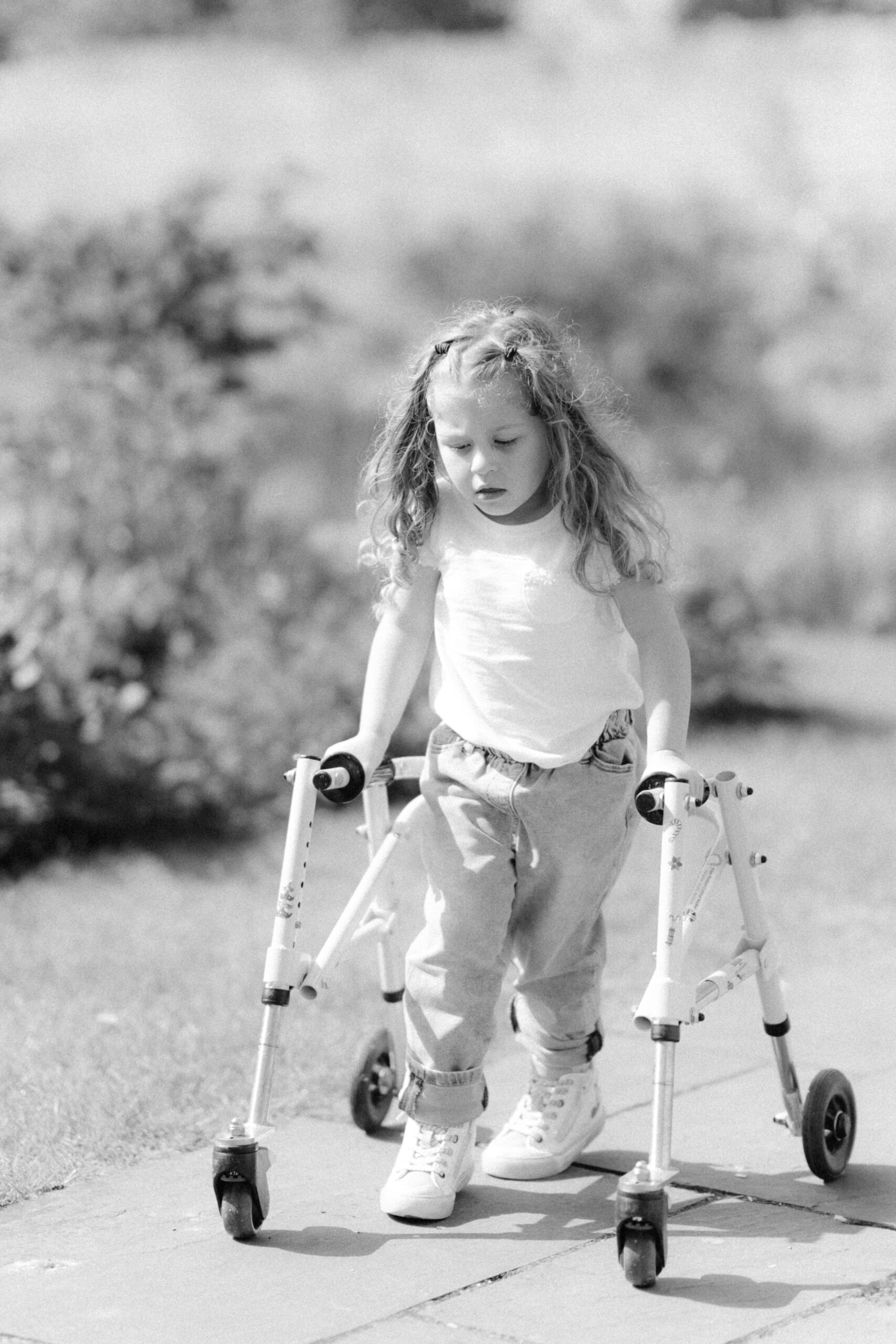
At Encephalon, I have the privilege of working with individuals and families navigating the profound challenges of neurological injury. One of the most striking cases I am beginning to work with is that of a four-year-old girl who suffered a stroke that resulted in paralysis of the left side of her body. Her story is both heart-wrenching and hopeful, as it reflects the resilience of the developing brain and the potential for targeted interventions.
This young child had already been diagnosed with cerebral palsy and faced significant challenges with walking. Prior to the stroke, she was making progress toward greater independence. However, the stroke caused a sharp decline, taking away much of the mobility she had achieved. She is now in the process of relearning how to walk, regaining fine motor control—especially in her left hand—and managing new layers of difficulty. Alongside motor challenges, she struggles with emotional dysregulation, expressed in severe tantrums and emotional outbursts that often trigger full-body stiffness. Compounding these challenges is significant fatigue, requiring frequent breaks throughout the day.
Why the Left Side Was Affected
The paralysis of her left side stems from damage to the right hemisphere of her brain, where the stroke occurred. The brain’s motor pathways cross over in the brainstem, meaning the right hemisphere controls movement and sensation on the left side of the body. The neurologist identified significant damage in the basal ganglia, a deep brain structure central to initiating and coordinating conscious movement. When the basal ganglia are impaired, an individual may experience both difficulties in fine motor skills and involuntary stiffness or rigidity.
The Basal Ganglia and Movement
The basal ganglia are not a single structure but rather a group of interconnected nuclei—including the caudate nucleus, putamen, and globus pallidus—that work in concert with the thalamus and cortex. Their primary role is to regulate voluntary movement by filtering out unnecessary motor activity and allowing purposeful, goal-directed actions to proceed smoothly. They act like a “gatekeeper” for movement: facilitating intended motor programs while inhibiting competing ones. Damage here disrupts the delicate balance between excitation and inhibition, leading to symptoms such as rigidity, slowed movements (bradykinesia), or involuntary movements. For this child, the stroke-induced damage to the basal ganglia likely contributes to her difficulties initiating and sustaining motor activity, as well as the episodes of stiffness triggered by emotional outbursts.
The basal ganglia also connect with emotional and cognitive networks. This overlap helps explain why damage in this region can influence both physical movement and emotional regulation simultaneously.
Emotional Dysregulation and the Right Hemisphere
Beyond motor function, the right hemisphere plays an essential role in emotional regulation and social perception. Damage here often results in difficulties with emotional processing, self-regulation, and behaviour. Networks involving the prefrontal cortex, limbic system, and basal ganglia are disrupted, leading to sudden mood shifts, frustration, and outbursts. For this child, the intertwining of emotional dysregulation and physical stiffness highlights the inseparability of mind and body in brain injury recovery.
The Role of Inflammation
Another critical factor in this case is neuroinflammation. Brain injury often sparks inflammatory processes that contribute not only to physical impairments but also to emotional and cognitive symptoms. Following a stroke, the body releases pro-inflammatory cytokines and activates microglial cells in an attempt to clear damaged tissue. While this is part of the healing process, chronic or excessive inflammation can impair recovery. It disrupts synaptic function, reduces neuroplasticity, and alters neurotransmitter balance (Brouns & De Deyn, 2009).
Research increasingly shows that inflammation can exacerbate fatigue, impair neuronal communication, and slow recovery (Woodburn et al., 2021). Moreover, neuroinflammation in the basal ganglia is linked with worsened motor symptoms, while inflammation in cortical-limbic networks can intensify emotional instability. Reducing inflammation, therefore, is not merely about easing discomfort—it is a central component of restoring optimal brain function and supporting emotional balance.
Linking Blood Pressure, Inflammation, and Neurofeedback
In my research experience, I have also observed how self-regulation practices can influence systemic physiology. For instance, using biofeedback, I measured blood pressure before and after a neurofeedback session and found a reduction in previously elevated levels. This is highly relevant, as hypertension is strongly associated with systemic inflammation and vascular dysfunction (Harrison et al., 2011). When nervous system arousal is reduced, not only can cardiovascular markers like blood pressure improve, but underlying inflammatory responses may also become less pronounced. This further highlights how interventions such as ILF Neurofeedback, which target regulatory networks in the brain, may help modulate not only neurological but also physiological dimensions of recovery.
My Approach: Stabilisation, Arousal, Mobility Through ILF Neurofeedback
My therapeutic focus will begin with stabilising the child’s nervous system using Infra-Low Frequency (ILF) Neurofeedback. This modality works by training the brain’s self-regulatory networks at a fundamental level, helping to improve stability, reduce hyperarousal, and restore a sense of balance. The initial goals are threefold:
- Reduce Inflammation: By supporting regulatory networks, ILF Neurofeedback may indirectly help mitigate neuroinflammatory responses, creating better conditions for recovery.
- Improve Emotional Regulation and Reduce Fatigue: By strengthening networks in the right hemisphere and basal ganglia, neurofeedback can support calmer emotional responses and reduce the energy expenditure associated with constant dysregulation.
- Target the Sensorimotor Strip: As the child progresses, training will focus more specifically on the sensorimotor cortex to enhance both global functioning (coordination, endurance) and localised control of the left side of the body.
Reflection
This case reminds me of the complexity of brain injury and the importance of tailoring intervention to both physiological and emotional needs. It also highlights the remarkable plasticity of the child’s brain, which, even after stroke, retains a profound capacity to reorganise and heal. With a steady focus on nervous system regulation, inflammation reduction, and targeted functional training, I am hopeful that we can support this child’s recovery journey in a meaningful way.
References
Brouns, R., & De Deyn, P. P. (2009). The complexity of neurobiological processes in acute ischemic stroke. Clinical Neurology and Neurosurgery, 111(6), 483–495.
Harrison, D. G., Guzik, T. J., Lob, H. E., Madhur, M. S., Marvar, P. J., Thabet, S. R., … & Weyand, C. M. (2011). Inflammation, immunity, and hypertension. Hypertension, 57(2), 132–140.
Woodburn, S. C., Bollinger, J. L., Wohleb, E. S. (2021). The semantics of neuroinflammation: What impact does it have on the brain and behavior? Frontiers in Behavioral Neuroscience, 15, 760.
Fisher, A. P., Reynolds, B. A. (2020). Potential of neurofeedback in the treatment of traumatic brain injury. Frontiers in Human Neuroscience, 14, 566.
Huster, R. J., Mokom, Z. N., Enriquez-Geppert, S., Herrmann, C. S. (2014). Brain–computer interfaces for EEG neurofeedback: Potentials and challenges. Frontiers in Neuroscience, 8, 369.
Marzbani, H., Marateb, H. R., Mansourian, M. (2017). Methodological note: EEG neurofeedback for psychological and neural rehabilitation – Principles and promise. NeuroImage: Clinical, 13, 465–474.